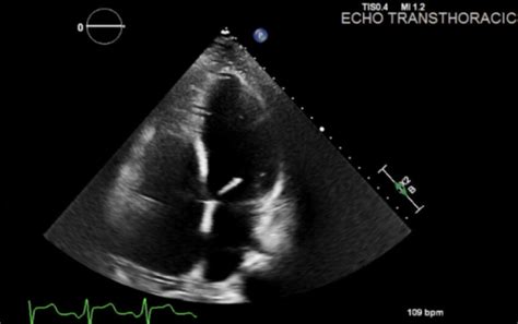
lv dilatation | cardiomegaly with biventricular dilatation lv dilatation Dilated cardiomyopathy affects the heart's ventricles and atria, the lower and upper chambers of the heart. The American Heart Association explains dilated . $36K+
0 · mildly dilated left ventricle symptoms
1 · left ventricle is severely dilated
2 · does dilated cardiomyopathy go away
3 · dilated cardiomyopathy signs and symptoms
4 · dilated Lv means
5 · causes of left ventricular dilation
6 · cardiomegaly with biventricular dilatation
7 · Lv dilatation definition
It all adds up to one thing: versatility. As shown here the classic model is 41mm with a teak-inspired horizontal dial pattern, but we particularly like the Aqua Terra in 38mm with a toned-down dial pattern. .
mildly dilated left ventricle symptoms
Dilated cardiomyopathy causes your heart’s main pumping chamber to expand. This decreases its ability to pump blood out to your body, putting you at risk for heart failure. The condition affects . The parasternal long axis and apical four-chamber views on transthoracic echocardiography are often the primary views used to gain both a qualitative and quantitative appreciation of left ventricular enlargement. . Tests to diagnose dilated cardiomyopathy include: Echocardiogram. This is the main test for diagnosing dilated cardiomyopathy. Sound waves produce images of the heart in motion. An echocardiogram .
Dilated cardiomyopathy affects the heart's ventricles and atria, the lower and upper chambers of the heart. The American Heart Association explains dilated .
left ventricle is severely dilated
does dilated cardiomyopathy go away
Dilated cardiomyopathy, or DCM, is when the heart chambers enlarge and lose their ability to contract. It often starts in the left ventricle (bottom chamber). As the disease gets worse, it may . Dilated cardiomyopathy (DCM) is characterized by dilation and impaired contraction of one or both ventricles [1-5]. Affected patients have impaired systolic function and . Dilated cardiomyopathy (DCM) is a heart condition in which the left ventricle weakens, stretches, and becomes larger, making it harder for the heart to pump effectively.
Dilated cardiomyopathy (DCM) is a heart muscle disease characterized by left ventricular or biventricular dilatation or systolic dysfunction without either pressure or volume .
Dilated cardiomyopathy is conventionally defined as the presence of left ventricular or biventricular dilatation or systolic dysfunction in the absence of abnormal loading conditions . Dilated cardiomyopathy is a type of heart muscle disease that causes the heart chambers (ventricles) to thin and stretch, growing larger. It typically starts in the heart's main pumping chamber (left ventricle).Dilated cardiomyopathy causes your heart’s main pumping chamber to expand. This decreases its ability to pump blood out to your body, putting you at risk for heart failure. The condition affects each person differently.
The parasternal long axis and apical four-chamber views on transthoracic echocardiography are often the primary views used to gain both a qualitative and quantitative appreciation of left ventricular enlargement. Features include 4: increased left ventricular internal end-diastolic diameter (LVIDd) Tests to diagnose dilated cardiomyopathy include: Echocardiogram. This is the main test for diagnosing dilated cardiomyopathy. Sound waves produce images of the heart in motion. An echocardiogram shows how blood moves in and out of the heart and heart valves. It can tell if the left ventricle is enlarged.
adidas neo herren sneaker schwarz
Dilated cardiomyopathy affects the heart's ventricles and atria, the lower and upper chambers of the heart. The American Heart Association explains dilated cardiomyopathy and the potential causes of dilated cardiomyopathy.
Dilated cardiomyopathy, or DCM, is when the heart chambers enlarge and lose their ability to contract. It often starts in the left ventricle (bottom chamber). As the disease gets worse, it may spread to the right ventricle and to the atria (top chambers). Dilated cardiomyopathy (DCM) is characterized by dilation and impaired contraction of one or both ventricles [1-5]. Affected patients have impaired systolic function and may or may not develop overt heart failure (HF). Dilated cardiomyopathy (DCM) is a heart condition in which the left ventricle weakens, stretches, and becomes larger, making it harder for the heart to pump effectively. Dilated cardiomyopathy (DCM) is a heart muscle disease characterized by left ventricular or biventricular dilatation or systolic dysfunction without either pressure or volume overload or coronary artery disease sufficient to explain the dysfunction.
Dilated cardiomyopathy is conventionally defined as the presence of left ventricular or biventricular dilatation or systolic dysfunction in the absence of abnormal loading conditions (eg, primary valve disease) or significant coronary artery disease sufficient to . Dilated cardiomyopathy is a type of heart muscle disease that causes the heart chambers (ventricles) to thin and stretch, growing larger. It typically starts in the heart's main pumping chamber (left ventricle).
Dilated cardiomyopathy causes your heart’s main pumping chamber to expand. This decreases its ability to pump blood out to your body, putting you at risk for heart failure. The condition affects each person differently. The parasternal long axis and apical four-chamber views on transthoracic echocardiography are often the primary views used to gain both a qualitative and quantitative appreciation of left ventricular enlargement. Features include 4: increased left ventricular internal end-diastolic diameter (LVIDd) Tests to diagnose dilated cardiomyopathy include: Echocardiogram. This is the main test for diagnosing dilated cardiomyopathy. Sound waves produce images of the heart in motion. An echocardiogram shows how blood moves in and out of the heart and heart valves. It can tell if the left ventricle is enlarged.

Dilated cardiomyopathy affects the heart's ventricles and atria, the lower and upper chambers of the heart. The American Heart Association explains dilated cardiomyopathy and the potential causes of dilated cardiomyopathy.
Dilated cardiomyopathy, or DCM, is when the heart chambers enlarge and lose their ability to contract. It often starts in the left ventricle (bottom chamber). As the disease gets worse, it may spread to the right ventricle and to the atria (top chambers).
Dilated cardiomyopathy (DCM) is characterized by dilation and impaired contraction of one or both ventricles [1-5]. Affected patients have impaired systolic function and may or may not develop overt heart failure (HF). Dilated cardiomyopathy (DCM) is a heart condition in which the left ventricle weakens, stretches, and becomes larger, making it harder for the heart to pump effectively.
Dilated cardiomyopathy (DCM) is a heart muscle disease characterized by left ventricular or biventricular dilatation or systolic dysfunction without either pressure or volume overload or coronary artery disease sufficient to explain the dysfunction.
dilated cardiomyopathy signs and symptoms
5, rue de Malte; 75011 Paris - France; Tel: +33 1 48 07 10 10; Fax: +33 1 48 05 90 17; [email protected]; Quick Menu. Special Offers; Photo Gallery; Location; Contact; .
lv dilatation|cardiomegaly with biventricular dilatation


